24
May

MicroRNA loaded extracellular vesicles affect tumor microenvironment in Tumor on a chip

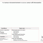
An interesting paper from Kim and Rhee groups at Korea Basic Science Institute and Incheon National University co-culturing glioblastoma with microglial cells in a tumor on a chip. They investigated therapeutic effects of microRNAs on tumor progression in brain cancer. The authors loaded microRNA-124 into extracellular vesicles, as a potential therapeutic agent, to regulate the crosstalk between tumor and microglia in the tumor microenvironment

Results
In a side-by-side channel chip, the authors co-cultured tumor and immune cells (microglia) close to each other (see methods below). Treatment with the microRNA inhibited the invasion of both cell types into a gel in the central channel, and resulted in more rounded, less invasive morphology in both tumor and microglial cells. Cytokine profiles also showed that after the treatment, most tumor progression-related cytokines were down-regulated except those associated with polarization of the microglia cells to M1 phenotype. Moreover, the treatment recruited more NK cells from the recirculation into the tumor microenvironment. This treatment has shown promising anti-tumor effects by decreasing related tumor markers, improving NK cell infiltration, and inhibiting microglial polarization.

2D mono-layer, Antibody microarray, Brain cancer, Cancer invasion, Cells suspended in hydrogel, Collagen type I, Efficacy, End-point microscopy, Immortalized cell line, miRNA therapeutics, On-chip monitoring, PDMS, Plasma treatment, Primary cells, Side-by-side channel chip, Soft lithography, Sterilization, Tumor immunology, Tumor microenvironment, Tumor on a chip, Tumor progression
Digestible of the paper Hong S, You JY, Paek K, Park J, Kang SJ, Han EH, Choi N, Chung S, Rhee WJ, Kim JA. Inhibition of tumor progression and M2 microglial polarization by extracellular vesicle-mediated microRNA-124 in a 3D microfluidic glioblastoma microenvironment. Theranostics 2021; 11(19):9687-9704. doi: 10.7150/thno.60851.Moreo. Link to paper: https://www.thno.org/v11p9687 This paper is reproduced under https://creativecommons.org/licenses/by/4.0/. The image of the chip was edited for better clarity, data in the table and text were compiled and interpreted by AZAR Innovations.

Methods
The authors used human glioblastoma cell line U373MG and immortalized SV40 microglia cell line (primary derived human microglia). MicroRNA-124 was loaded into HEK293T-derived extracellular vesicles which were then injected to the side channels. 
Fabrication: Soft lithography with PDMS
Sterilization: Plasma treatment (day -1)
Cell incorporation: 
Collagen type I hydrogel was injected to a side-by-side channel chip, and the cells were either cultured in the two side channels (on the side wall of the gel) or in 3D in the gel (day0). Natural killer cells were added to the side channels (day 2).
Perfusion/refreshing: The media in the cell channels were replaced every day (day 0-4).
Treatment: MicroRNA-loaded extracellular vesicles were loaded to the side channels (day 0-4).

On-chip read-outs: End-point microscopy (bright field and fluorescence, day 2 & 4)
Off-chip read-outs: Cytokine profiling (Antibody microarray)


Strong points:
+ Investigation of different microRNA anti-tumor effects in different cell lines.
+ Differen
t methods of microRNA delivery into cells.
+ Co-culture of glioblastoma cells with different immune cells

Negative points:
– The same old story, they used PDMS but performed no investigation of the extracellular vesicle penetration into PDMS.
– They had to refresh the media in the chips manually everyday although the co-culture was for 4 days.

Conclusion and outlook
The authors used a microfluidic system to better understand the glioblastoma microenvironment involving tumor and immune cells. This platform can be used to develop potential therapeutic strategies using miRNA-loaded extracellular vesicles.


Have any questions? Leave a comment below or contact us.