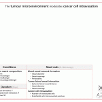
Matrix composition modulates vasculature formation and intravasation in tumor chip
This is a common question: what extracellular matrix (ECM) should I use? Specifically, the choice of matrix is very important to mimic the tumor–stromal cells interaction during cancer intravasation. This paper by Agrawal et al. from the Moeendarbary lab at University College London uses a tumor on a chip to study the effect of different ECM compositions on vasculature formation and cancer cell intravasation.
Results
The authors first studied the effect of different matrices on size and coverage area of the vessels. After adding tumor spheroids, the distance between the vessels and the spheroids increased significantly when they were cultured in fibrin only, meaning that adding Matrigel and collagen type I enhanced the contact between the vessels and the tumor. The presence of tumor spheroid decreased the average vessel diameter. Vessel coverage area was larger, with and without tumor, when cultured in the hydrogel with all the matrix components. Interestingly, cancer cell intravasation occurred only where all the matrix components were present.

Methods
The authors used a 3-lane side-by-side chip, added the spheroids (made using human colorectal adenocarcinoma HT29 and human lung cancer cell line A549 in U-bottom plates), the extracellular matrix (fibrin, fibrin:collagen type I, fibrin:Matrigel, fibrin:collagen type I:Matrigel), endothelial cells (HUVECs) and fibroblasts (normal human lung fibroblasts).
Fabrication: laser cutting and 3D printing the mold, soft lithography with PDMS
Sterilization: PDMS device autoclaved, then bonded to glass with plasma, then put at 80˚C to restore hydrophobicity
Cell incorporation: Cells and matrices injected via the inlet hole into the central channel
Perfusion/refreshing method: Manual medium exchange every 24 hours
On-chip read-outs: Immunostaining, vessel perfusion with 70 kDa fluorescent dextran (injection into one side channel only), end point and real-time fluorescent microscopy
Improvement points
– Use of other materials than PDMS
– More automatic instead of manual refreshing of medium
– More automatic image analysis
Strong points:
+ Different matrix compositions
+ Tested with different cancer types
+ Real-time imaging
In summary:
A beautiful work emphasizing the importance of choosing the right extracellular matrix as the tumor microenvironment in a tumor on a chip. This technology enables researchers to study the effect of tumor microenvironment on cancer metastasis more mechanistically.
Do you have questions about the system in this paper or similar technologies? Or do you want to start with organ on a chip? Feel free to contact us!