Platelet-tumor interaction in ovarian cancer metastasis
A recent paper from Jain lab at Texas A&M University that studies the extravasation of platelets into the ovarian tumor microenvironment in a microfluidic chip. The presented tumor-on-a-chip reveals the dynamics of cancer cell-platelet interaction towards more metastatic behavior in cancer cells.
Results
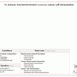
The authors published a similar technology in their previous paper to study the dynamics of platelet extravasation. The previous system comprised only two microchannels on top of each other separated by a PDMS membrane. This time, they added more complexity to the system to study the 3D invasion of cancer cells into an ECM by adding two compartments in the top layer (Similar to the chips developed by Roger Kamm). The results indicated that platelets and tumors interact under shear through glycoprotein VI (GPVI) and tumor galectin-3.

DigesTable of the paper Saha, B., et al. (2021). “Human tumor microenvironment chip evaluates the consequences of platelet extravasation and combinatorial antitumor-antiplatelet therapy in ovarian cancer.” Science Advances 7(30): eabg5283. This paper is reproduced under https://creativecommons.org/licenses/by/4.0/. The image of the chip was edited for better clarity, data in the table and text were compiled and interpreted by AZAR Innovations.
Method
The authors cultured human ovarian microvascular endothelial cells (HOMEC) and ovarian carcinoma A2780 cells. Platelets were isolated from fresh human donor blood samples. They used a hybrid design chip.
Fabrication: soft lithography with PDMS
Sterilization
Cell incorporation: HOMEC cell suspension was injected inside the vessel chamber and cancer cell suspension was seeded on the upper chamber
Perfusion/refreshing: they used a syringe pump to perfuse the platelets for one day into the vascular channel and later to induce shear stress on the ovarian cancer cells in the top compartment.
Treatment: Cisplatin and Revacept
On-chip read-outs: end-point microscopy
Off-chip read-outs: off-chip imaging, immuno-histo chemistry, Flow cytometry, RNA-seq
Strong points:
+ Measure the physical interaction between cancer cells and platelets
+ Controlled compartmentalization of the chip
+ Extensive RNA-Seq analysis of the cells isolated from the chip
Nothing is perfect! The system can also improve:
– No analysis of absorption of the drugs into PDMS
– Although cancer cells invade into a 3D ECM gel, they were cultured in 2D
– They used a syringe pump to perfuse the chip which makes the system’s scale up difficult and increases the media consumption.
Conclusion and outlook
This tumor on a chip model can be used to study the interactions between blood cells and cancer cells. It can also play a significant role in drug development and anti metastasis drugs investigations.
Contact us if you want to know more about this system or similar technologies!


